September 6, 2024
Ultrasound Physical Rehabilitation: Benefits, Adverse Effects & Treatments
Ultrasound Physical Rehabilitation: Advantages, Side Effects & Therapies Core temperatures of experimental animals are different from those of people, and extrapolation of thermal injury information from pet designs to humans may be difficult. A lot of researches linking the potential neonatal impacts of ultrasound have actually not been Body Contouring consistently validated. For example, restriction required for ultrasound exam in the conscious animals is a known teratogen.152 The visibility of unacknowledged maternal or congenital condition or toxic substance direct exposure may likewise confuse analysis of research studies of ultrasound biologic effects.Biological Effects Of Ultrasound
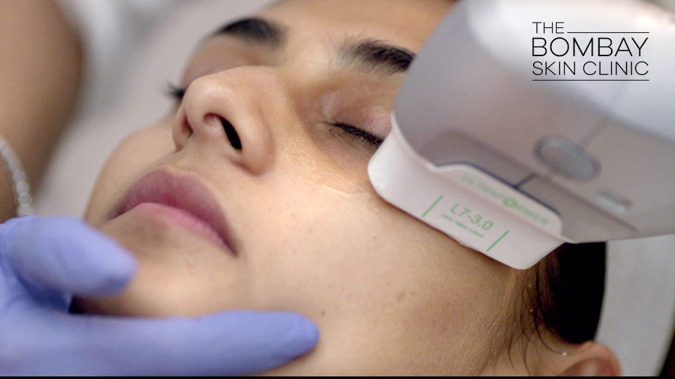
By Brett Sears, PTBrett Sears, PT, MDT, is a physiotherapist with over 20 years of experience in orthopedic and hospital-based treatment. 2 review authors separately analyzed the threat of bias of each test and drawn out the data. We did a meta-analysis when sufficient professional and analytical homogeneity existed. We established the certainty of the evidence for each and every contrast utilizing the quality approach. Research studies have shown scientifically substantial improvement in eyebrow lift and in skin laxity of the reduced face and neck, with high patient-reported satisfaction.Us Fda
- Lithotripsy is likewise accomplished by minimally intrusive probes which are advanced to the stone.
- Nonetheless, the moderate-temperature hyperthermia method has actually not progressed to extensive medical usage, and the effort in hyperthermia cancer cells therapy has shifted to using high strength focused ultrasound.
- If you are experiencing discomfort or a current injury, ultrasound therapies may benefit you.
- The Globe Federation of Ultrasound in Medicine and Biology and the European Federation of Societies for Ultrasound in Medication and Biology further suggest limiting the ultrasound direct exposure duration during Doppler mode sonography (fig. 4).
What Are The Various Sorts Of Ultrasound Therapy Treatments?
Does ultrasound recover nerves?
Luckily, previous studies have actually revealed that low-intensity pulsed ultrasound (LIPUS) has the potential to induce nerve regrowth by promoting neurotrophic variables and reducing neuroinflammation.
Social Links